
Echocardiography

This ultrasound examination shows the heart chambers, valves and pumping function in real time - painless and without radiation.
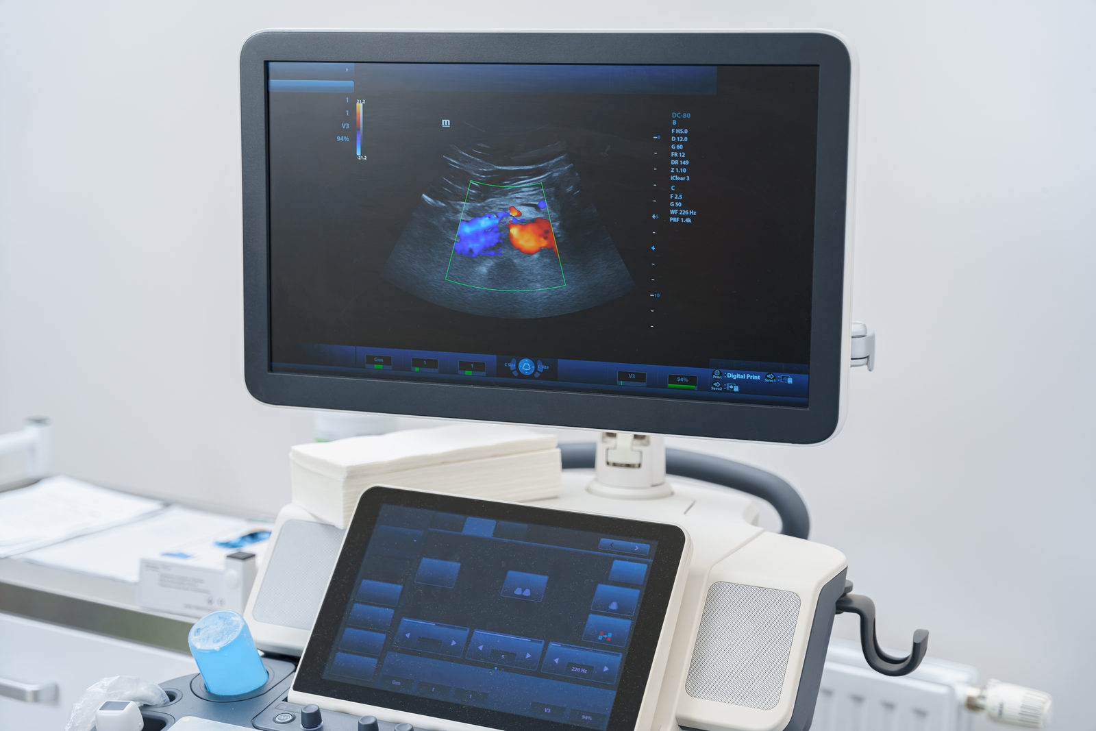
Ultrasound machine showing a colour Doppler image

Seeing the heart in motion

A probe placed on the outside of your chest produces moving images of your heart. This allows us to assess the size of the heart chambers, the thickness of the heart muscle, the pumping function and how well the heart valves work.

Doppler technology also makes the blood flow inside the heart visible. This helps detect, for example, leaking or narrowed heart valves.

When is echocardiography useful?

  • A heart murmur heard with a stethoscope
  • Shortness of breath or reduced fitness
  • Swelling in the legs
  • Long-standing high blood pressure
  • Suspected heart failure or heart valve disease
  • Follow-up of a known heart condition

In case of acute chest pain, shortness of breath or loss of consciousness, call the emergency number 112 immediately.

What happens during echocardiography

  • 01
    PositioningYou undress to the waist and lie on your left side.
  • 02
    Ultrasound gelA gel ensures the sound waves are transmitted well.
  • 03
    ExaminationThe probe is placed at different points on your chest. This usually takes about 20 to 30 minutes.
  • 04
    DiscussionYou find out straight afterwards what the images show.

How to prepare

  • No special preparation needed
  • You do not need to fast
  • Take your medication as usual
  • Wear comfortable clothing that is easy to open
  • Bring previous test results, if available

Related services

Your doctor

Book an appointment

Book online via Doctolib or call us during consultation hours.