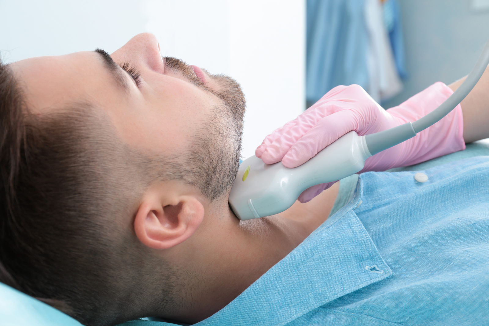
What does vascular ultrasound show?
Doppler and duplex ultrasound show the carotid arteries and other blood vessels in the neck. The images reveal the vessel wall as well as possible deposits (plaques) and calcification.
Doppler technology also measures the speed of the blood flow. This makes it possible to detect narrowing and assess how severe it is. The examination is painless and does not use X-rays.
When is vascular ultrasound useful?
- Several risk factors such as high blood pressure, diabetes, raised cholesterol or smoking
- A known vascular condition, for example coronary artery disease or poor circulation in the legs
- An unusual flow sound over the carotid artery
- Follow-up of known narrowing or deposits
In case of sudden paralysis, speech or vision problems, call the emergency number 112 immediately - these can be signs of a stroke.
What happens during the examination
- 01
PositioningYou lie comfortably on your back with your neck uncovered.
- 02
Ultrasound gelA gel ensures the sound waves are transmitted well.
- 03
ExaminationThe probe is moved along the side of your neck. This usually takes about 15 to 20 minutes.
- 04
DiscussionYou find out straight afterwards what the images show.
How to prepare
- No special preparation needed
- You do not need to fast
- Take your medication as usual
- Wear clothing with an open neckline and remove necklaces
- Bring previous test results, if available
Your doctor

Dr. medic Stergios Chantzaras
Specialist in Internal Medicine and Cardiology
View profile