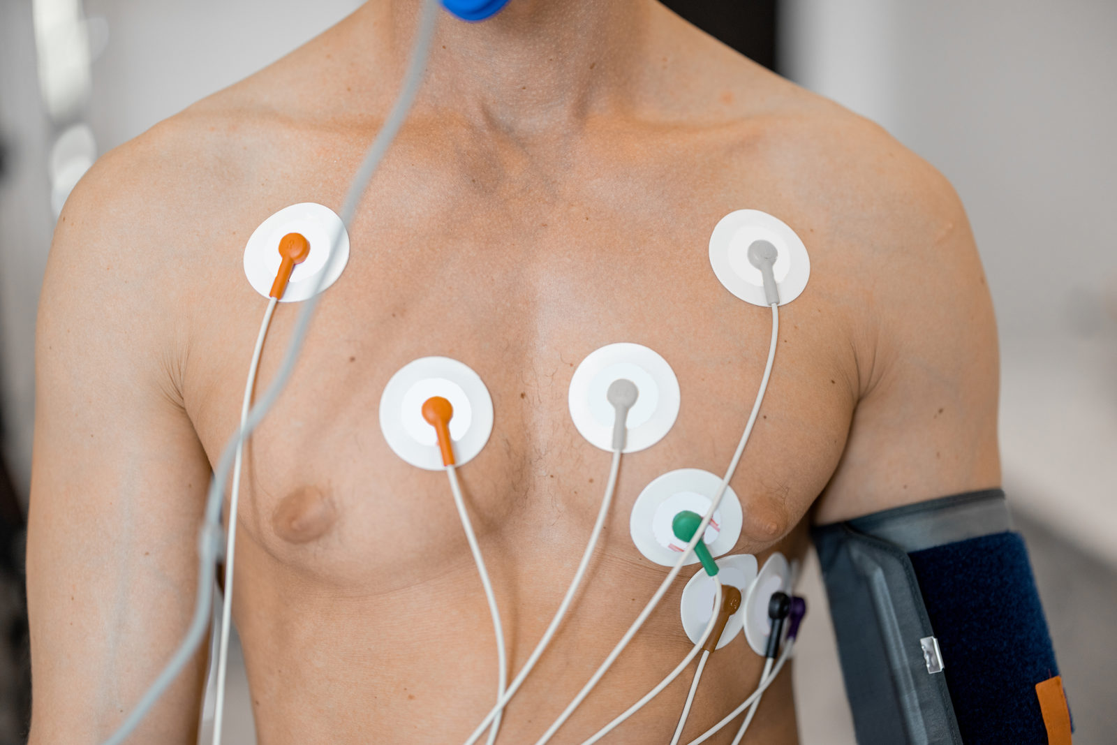
How does your heart work under exertion?
Some heart conditions only become apparent when the heart has to work harder. Stress echocardiography therefore compares ultrasound images of the heart at rest with images taken under exertion. This shows whether all areas of the heart muscle move evenly and receive enough blood during effort.
The test does not use X-rays. Your heart rhythm and blood pressure are monitored throughout the examination.
When is stress echocardiography useful?
- Suspected reduced blood flow in the coronary arteries (coronary artery disease)
- Chest tightness or shortness of breath during physical activity
- Follow-up of known coronary artery disease
- Assessment of certain heart valve conditions under exertion
- An unclear exercise ECG result
In case of acute chest pain, shortness of breath or loss of consciousness, call the emergency number 112 immediately.
What happens during stress echocardiography
- 01
PreparationYou are connected to an ECG and blood pressure monitor.
- 02
Images at restFirst, your heart is examined with ultrasound at rest.
- 03
ExertionThe workload is increased step by step, for example on a recumbent ergometer, while your heart continues to be observed with ultrasound. You can report any symptoms at any time.
- 04
Recovery and discussionAfter a short recovery phase, we discuss the result with you. Please allow about 45 minutes in total.
How to prepare
- Wear comfortable sportswear and sturdy shoes
- Eat only a light meal beforehand
- We will discuss with you whether medication such as beta blockers should be paused - please do not stop any medication on your own
- Bring your current medication plan and previous results
- If you have a fever or an acute infection, please reschedule your appointment
Your doctor

Dr. medic Stergios Chantzaras
Specialist in Internal Medicine and Cardiology
View profile